A microscope that has been a key research tool for students and faculty at the University of Hawaiʻi at Mānoa, as well as the broader Hawaiʻi science community, is nearing the end of its life, and researchers are searching for its replacement.
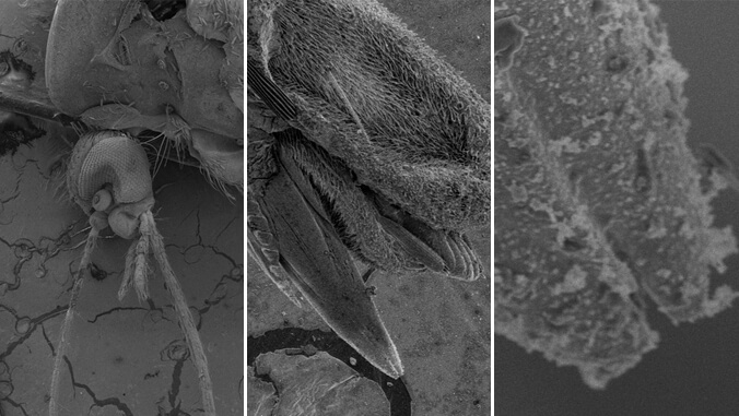
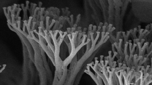
The scanning electron microscope (SEM) has been a facet in UH Mānoa’s Biological Electron Microscope Facility (BEMF) since 2009. It has been used by more than 400 researchers. SEM works by using a beam of electrons scanned over the surface of a sample to get images of that surface. Electrons get a better resolution than photons (light), so extremely small details can be observed. A light microscope can show tissues and cells, however, an electron microscope is able to see things much smaller, such as bacteria and other pathogens, as well as the ultra fine details of biological and engineered surfaces.
“A new SEM will increase research capacity, and support UH Mānoa faculty in attracting extramural funds,” BEMF Director Alex Culley said. “The need for the new microscope is immediate. It is essential in order to avoid significant disruption to the research programs of the more than 65 users who are dependent on the instrument. It is also necessary to ensure that a functional SEM is available to the students who receive training as part of the core curriculum in the biological sciences.”
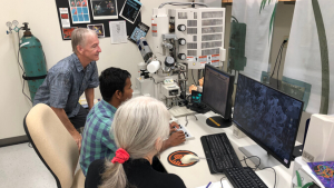
According to BEMF Lab Supervisor Tina Weatherby Carvalho, as the microscope ages, the quality of its performance declines. SEM was purchased through grant funding in January 2009 for just over $437,000. Microscopes of this nature generally last around 10 years. Its longer life is credited to Carvalho and her team who have performed regular maintenance. Once the microscope is unable to perform its duties, the team will look to purchase a replacement, which is estimated to cost around $650,000 today. One that is fully-equipped can cost upwards of $900,000.
“Think of some of these high-tech instruments as you would an automobile,” Carvalho said. “When they are new, they have the latest technology, have been through rigorous testing and their performance is top-notch. As they age, performance declines until eventually they don’t meet specifications. Parts become difficult to get and support gets spotty. And as years pass users start to ask for the newest features. UH researchers acquire funding based on grant proposals where they have to specify what kind of data they will be able to acquire, and we have to continue supporting that.”
Although replacing and upgrading the SEM is one of the BEMF’s primary challenges, the facility has recently increased its capacity to meet the needs of the university and beyond.
“We have added and upgraded instruments, brought in new personnel and continue to offer new services,” Culley said. “We have added a multi-spectral confocal laser scanning microscope which produces incredibly high resolution images of live samples, among other capabilities. With support from the U.S. Geological Survey, the transmission electron microscope now features a cooled charged-coupled device camera that has dramatically increased the sensitivity of the instrument. We are also fortunate enough to have recently hired Kris Ewell. Kris has decades of experience in histology and pathology and now brings these skills to the BEMF.”
Invaluable research tool
SEM is one of several different types of specialized microscopes in BEMF. All users pay an hourly fee to use the microscope, with a discount for UH-affiliated researchers.
“SEM is a foundational tool and at UH Mānoa it is used by researchers in the life and biomedical sciences, oceanography, engineering, materials science and others,” Culley said. “Current areas of biological research that require ultra-high resolution scanning electron microscopy include microbiology, pathogen-host studies, biodiversity, sensory and cell biology, and neuroscience. SEM is an important tool in non-biological applications, such as in the production of microelectronics, semiconductors, and even forensic investigations.”
UH Mānoa School of Life Sciences Professor Les Watling said the SEM has been indispensable to his research, and uses the microscope to get very clear, wide depth of focus images of small crustaceans or deep sea corals, the two groups he studies for taxonomy and functional anatomy.
“Over the years I have spent many hours using that SEM and I am sure I have thousands of images on my computer that have been taken using it,” Watling said. “Some of the images are truly utilitarian, that is, used for descriptive or measurement purposes, but other times I see things that strike an artistic chord, so I try to spend a little time getting a truly ‘art’ shot, but one that can also be used for my scientific work.”
Dennis Kunkel is an expert in using SEM for research and was affiliated with two UH departments from 1993–2000. He has assisted former BEMF Director Richard Allen with studying how a one-celled organism digests its food as well as a unique mechanism for motility, and former Associate Researcher Jes Stollberg with developing frog muscle cells. In 2000, research funds were not available so Kunkel decided to work as an electron microscope consultant with professors, science teachers, other laboratories and businesses.
Kunkel’s award-winning images appear worldwide in print, film and digital media such as TIME magazine, Scientific American, The New York Times, Life and National Geographic. He has made significant contributions in the fields of microbiology, neurobiology and botany, and authored or co-authored 62 research papers in scientific journals.
“Electron microscopy, especially scanning electron microscopy, is a very valuable tool in many scientific areas and industries,” Kunkel said. “Since the BEMF is a user facility, I could continue with the SEM consulting and pay an outside user fee to UH to help support the facility. Being able to have a high quality SEM has allowed me to work on many biological subjects.”
BEMF is part of the Pacific Biosciences Research Center, which is housed in the School of Ocean and Earth Science and Technology.
—By Marc Arakaki