A Forearm Deformity in a 4 Year Old
Radiology Cases in Pediatric Emergency Medicine
Volume 6, Case 16
Loren G. Yamamoto, MD, MPH
Kapiolani Medical Center For Women And Children
University of Hawaii John A. Burns School of Medicine
This is a 4 year old female who presents to the
emergency department with a forearm injury after falling
off the jungle gym (playground bars) at the park. Her
mother noted that her forearm was deformed and she
was complaining of persistent pain. She denies trauma
or pain elsewhere.
Her past medical history is unremarkable.
Exam: VS are normal. She is alert and comfortable
in no distress. HEENT unremarkable. Chest and
abdomen unremarkable. There is a modest deformity
of her left mid-forearm. Tenderness is noted in the area
of the deformity. Her wrist and hand are non-tender.
No bruising is noted. Her pulses are good and
sensation is intact. She moves her fingers well. Her
elbow and humerus are non-tender.
Radiographs of her left forearm are obtained.
View forearm radiographs.
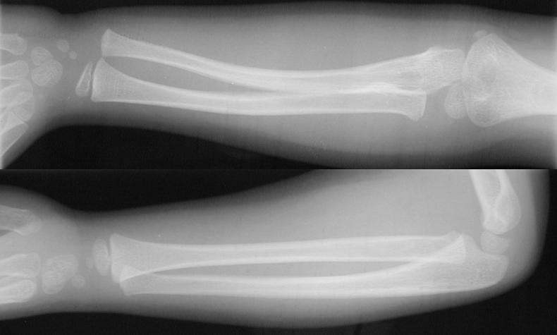 Although there is an obvious deformity of her
forearm on exam, no fracture is evident here. Her
elbow does not demonstrate a joint effusion and her
radial head is of normal contour and is well aligned with
the capitellum (refer to Case 18 of Volume 2, Test Your
Skill In Reading Pediatric Elbows).
Note the curvature of the ulna which is excessive.
This represents a "bowing fracture" of the ulna. Bowing
fractures usually occur in the forearm. This is a
bending deformity without a grossly visible fracture in
the tubular structure of the bone. Microfractures are
present on microscopy, but only the bowing is
appreciable on plain radiographs. Reduction of a
bowing fracture requires a lot of force, thus it should be
done under general anesthesia.
Failure to recognize a bowing fracture of the
forearm results in limited supination and pronation.
Periosteal reaction on later radiographs may not occur
with bowing fractures so this cannot be used as a
criterion to rule out an earlier fracture.
A comparison view of the other extremity may be
useful in identifying the bowing fracture.
View comparison of the other forearm.
Although there is an obvious deformity of her
forearm on exam, no fracture is evident here. Her
elbow does not demonstrate a joint effusion and her
radial head is of normal contour and is well aligned with
the capitellum (refer to Case 18 of Volume 2, Test Your
Skill In Reading Pediatric Elbows).
Note the curvature of the ulna which is excessive.
This represents a "bowing fracture" of the ulna. Bowing
fractures usually occur in the forearm. This is a
bending deformity without a grossly visible fracture in
the tubular structure of the bone. Microfractures are
present on microscopy, but only the bowing is
appreciable on plain radiographs. Reduction of a
bowing fracture requires a lot of force, thus it should be
done under general anesthesia.
Failure to recognize a bowing fracture of the
forearm results in limited supination and pronation.
Periosteal reaction on later radiographs may not occur
with bowing fractures so this cannot be used as a
criterion to rule out an earlier fracture.
A comparison view of the other extremity may be
useful in identifying the bowing fracture.
View comparison of the other forearm.
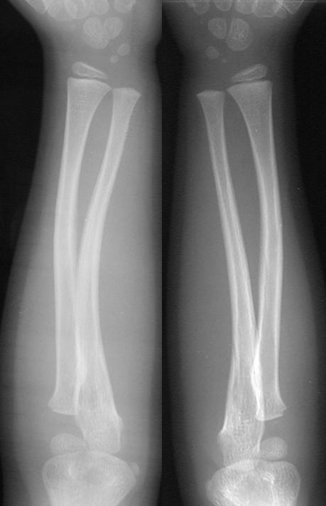 This comparison view shows the normal
configuration of the unaffected right radius and ulna on
the right image. Comparing this with her affected left
forearm (left image), it is easier to appreciate the
bowing deformity of the ulna. However, note that the
two are not very different since the bowing of the left
ulna is not severe.
Arrows point to the bowing deformity of the ulna.
This comparison view shows the normal
configuration of the unaffected right radius and ulna on
the right image. Comparing this with her affected left
forearm (left image), it is easier to appreciate the
bowing deformity of the ulna. However, note that the
two are not very different since the bowing of the left
ulna is not severe.
Arrows point to the bowing deformity of the ulna.
 Examine the left forearm in isolation.
See if you can appreciate the bowing deformity of the ulna.
In this case, the clinical appearance of a
deformed forearm is highly indicative of a
fracture. If radiographs fail to confirm the presence
of an obvious fracture, consider the possibility of a
bowing fracture.
References
Diaphysis (Chapter 16). In: Harris JH, Harris WH,
Novelline RA. The Radiology of Emergency Medicine,
third edition. 1993, Baltimore, MD, Williams & Wilkins,
pp. 1059-1061.
Examine the left forearm in isolation.
See if you can appreciate the bowing deformity of the ulna.
In this case, the clinical appearance of a
deformed forearm is highly indicative of a
fracture. If radiographs fail to confirm the presence
of an obvious fracture, consider the possibility of a
bowing fracture.
References
Diaphysis (Chapter 16). In: Harris JH, Harris WH,
Novelline RA. The Radiology of Emergency Medicine,
third edition. 1993, Baltimore, MD, Williams & Wilkins,
pp. 1059-1061.
Return to Radiology Cases In Ped Emerg Med Case Selection Page
Return to Univ. Hawaii Dept. Pediatrics Home Page