Abdominal/Hip Pain With Fever in a 2-Year Old
Radiology Cases in Pediatric Emergency Medicine
Volume 5, Case 12
Rodney B. Boychuk, MD
Kapiolani Medical Center For Women And Children
University of Hawaii John A. Burns School of Medicine
This 2-year old female was in her usual state of
good health until one month prior, when she began
complaining of generalized abdominal pain. The "pain
spasms" occurred every 15-20 minutes, which would
cause her to "double over"; the pain would then
gradually subside. At that time, she was seen in the
emergency department, where abdominal radiographs
revealed moderate gas throughout the bowel and a
stool-filled colon. An enema resulted in the passage of
large amounts of stool, and the pain seemed to
improve. About one week ago (three weeks later), her
pain became severe again and, at this point, she
refused to walk. Her parents noticed that her knees
would shake when she stood up, and she appeared
knock-kneed. For the last 3 days, these episodes have
intensified, with each episode lasting approximately 5
minutes and returning every 15-20 minutes. Her
temperature ranged from 37.3 to 38.6 degrees. She
was noted to be more agitated at night and was unable
to sleep because of pain. She also seemed to favor
her right leg. There was no nausea, vomiting, diarrhea,
cough, rhinorrhea or other symptomatology.
Exam: VS T40.3, P160, RR 46, BP 124/65. She
appears tired, but otherwise well-developed and
well-nourished. She is somewhat irritable but
cooperative. She is in the 50th percentile for height,
weight and head circumference. HEENT no
abnormalities detected. Neck supple without
adenopathy. Heart regular without murmurs. Lungs
clear. Abdomen is soft without definite tenderness.
Bowel sounds are active. No rebound. No
organomegaly. No hernias. Small 1 cm lymph nodes
are palpable in both inguinal regions. There is
tenderness to palpation in the right hip area. Range of
motion about this hip is good; however, she fusses
when this is done. There does not appear to be any
pain with palpation of the pubis, and she is able to log
roll normally. There are no other bony or joint
abnormalities noted.
Initial labs revealed a white count of 21,700 with
60% neutrophils, 6% bands, 25% lymphs, 8% monos,
and 1% eos. Hgb 12.3, Hct 37.2. Platelet count
295,000. Her ESR is 42. Chemistry panel shows a
slightly increased LDH at 382 and slightly increased
alkaline phosphatase at 212. Radiographs of the
abdomen and pelvis are ordered.
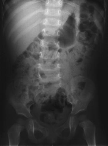
View abdominal flat plate.
 View pelvic radiographs.
 A previous abdominal series done one month ago
showed a lot of stool. The overall abdominal gas
pattern is non-specific and largely unchanged from the
previous radiographs one month ago.
The radiograph of the pelvis appears fairly normal at
first glance. Examine the bones carefully to see if you
can detect any abnormalities.
This pelvis shows cortical thickening and sclerosis of
the right superior pubic ramus. Compare the right and
the left sides. You can see that the right superior pubic
ramus is hyperdense and irregular. This finding can
also be appreciated in retrospect on the abdominal film
as well. The radiologist feels that this could be
secondary to a low-grade infection, the healing phase of
histiocytosis, or a malignancy.
A CT scan of the abdomen showed no other
abnormalities. It confirmed the slight cortical sclerosis
and thickening of the right superior pubic ramus seen
on the plain radiographs.
A radionuclide bone scan was ordered.
View bone scan.
A previous abdominal series done one month ago
showed a lot of stool. The overall abdominal gas
pattern is non-specific and largely unchanged from the
previous radiographs one month ago.
The radiograph of the pelvis appears fairly normal at
first glance. Examine the bones carefully to see if you
can detect any abnormalities.
This pelvis shows cortical thickening and sclerosis of
the right superior pubic ramus. Compare the right and
the left sides. You can see that the right superior pubic
ramus is hyperdense and irregular. This finding can
also be appreciated in retrospect on the abdominal film
as well. The radiologist feels that this could be
secondary to a low-grade infection, the healing phase of
histiocytosis, or a malignancy.
A CT scan of the abdomen showed no other
abnormalities. It confirmed the slight cortical sclerosis
and thickening of the right superior pubic ramus seen
on the plain radiographs.
A radionuclide bone scan was ordered.
View bone scan.
 There is intense uptake in the right superior pubic
ramus. The differential includes malignancy versus
infection. The L1 vertebral body is abnormal in that the
left lateral aspect of it appears to be "cold".
In the operating room, an open biopsy was sent
for histology and culture. A frozen section of curettings
was read as "possible tumor". The cultures returned
negative and the final histology was read as chronic
inflammation and fibrosis.
She was continued on antibiotics for suspected
osteomyelitis. However, she did not improve and her
fever worsened. A bone marrow aspiration was
performed which showed an unusual type of acute
leukemia.
Discussion
Skeletal changes that occur in leukemia are due to
infiltration of the bone by proliferating white cells.
Characteristically, zones of rarefaction with
subperiosteal new bone formation in the metaphyseal
region of the humerus or femur, or in the pelvis or
spine, are also seen. Occasionally, there is a
widespread, diffuse rarefaction of the skeleton (1).
Lucent metaphyseal bands are said to be
characteristic of leukemia; however, Rogalsky, et al (3)
found lytic lesions in 19%, sclerotic lesions in 4%, and
periosteal new bone formation in 2%.
In the younger child, an irregular lytic lesion and/or
the presence of periosteal new bone, with or without a
lytic lesion, should always suggest osteomyelitis,
metastatic neuroblastoma, and eosinophilic granuloma,
along with leukemia (6). In the older child and
adolescent, various forms of subacute osteomyelitis
most often mimic tumors (6).
Osteomyelitis is an inflammation of the bone.
Petrola and Vahvanem (5) consider the diagnosis
established when 2 of the 4 following criteria are
present: 1) Pus aspirated from bone; 2) Positive bone
or blood culture; 3) Classic symptoms of localized pain,
swelling, warmth, and limited range of motion of the
adjacent joint; and 4) Radiographic changes
characteristic of osteomyelitis.
When these criteria are not met, it is helpful to
remember those conditions that may mimic
osteomyelitis and therefore be mistaken for such (6).
Trauma may be the most common. It shares some
clinical features with osteomyelitis, including pain,
tenderness, swelling, and soft tissue swelling on
radiographs. Trauma pain improves with time, whereas
osteomyelitis worsens. Another differentiating feature
is the ESR, which is elevated with osteomyelitis, but
normal with trauma.
Neoplasms may mimic osteomyelitis. Leukemia is
the most common malignancy in childhood, with bone
pain being the presenting sign in about 30% (2). Other
non-specific symptoms, such as fever, lethargy, and an
elevated ESR and WBC are often present as well.
When considering leukemia, other signs and symptoms
must be aggressively sought: bone pain in multiple
sites, easy bruising, bleeding, a low white blood cell
count, anemia, thrombocytopenia, etc.
References:
1. Outline of Orthopedics, 11th ed., John Crawford
Adams David L. Hamblem, Churchill Livingston, 1990,
p. 91.
2. Hamm IM, Guppa S, Palmer MK, et al. The
prognostic significance of radiological and symptomatic
bone involvement in childhood acute lymphoblastic
leukemia. Med Pediatr Oncol 1979;6:51.
3. Rogalsky RJ, Black GB, Reed MH. Orthopedic
manifestations of leukemia in children. J Bone Joint
Surg (Am) 1986;68:494.
4. Clausen N, Gortz H, Petersen A, et al. Skeletal
scintigraphy and radiography at onset of acute
lymphoblastic leukemia in children. Med Pediatr Oncol
1983;11:291.
5. Ptrola H, Vahvanen V. A comparative study of
osteomyelitis and purulent arthritis with special
reference to etiology and recovery. Infection
1984;12:55.
6. Morrissy RT. Bone and Joint Sepsis. In:
Morrissy RT, Weistein SL (eds). Lovell and Winters
Pediatric Orthopedics, 4th ed. Lippincott-Raven
Publishers, 1996, pp. 579-619.
There is intense uptake in the right superior pubic
ramus. The differential includes malignancy versus
infection. The L1 vertebral body is abnormal in that the
left lateral aspect of it appears to be "cold".
In the operating room, an open biopsy was sent
for histology and culture. A frozen section of curettings
was read as "possible tumor". The cultures returned
negative and the final histology was read as chronic
inflammation and fibrosis.
She was continued on antibiotics for suspected
osteomyelitis. However, she did not improve and her
fever worsened. A bone marrow aspiration was
performed which showed an unusual type of acute
leukemia.
Discussion
Skeletal changes that occur in leukemia are due to
infiltration of the bone by proliferating white cells.
Characteristically, zones of rarefaction with
subperiosteal new bone formation in the metaphyseal
region of the humerus or femur, or in the pelvis or
spine, are also seen. Occasionally, there is a
widespread, diffuse rarefaction of the skeleton (1).
Lucent metaphyseal bands are said to be
characteristic of leukemia; however, Rogalsky, et al (3)
found lytic lesions in 19%, sclerotic lesions in 4%, and
periosteal new bone formation in 2%.
In the younger child, an irregular lytic lesion and/or
the presence of periosteal new bone, with or without a
lytic lesion, should always suggest osteomyelitis,
metastatic neuroblastoma, and eosinophilic granuloma,
along with leukemia (6). In the older child and
adolescent, various forms of subacute osteomyelitis
most often mimic tumors (6).
Osteomyelitis is an inflammation of the bone.
Petrola and Vahvanem (5) consider the diagnosis
established when 2 of the 4 following criteria are
present: 1) Pus aspirated from bone; 2) Positive bone
or blood culture; 3) Classic symptoms of localized pain,
swelling, warmth, and limited range of motion of the
adjacent joint; and 4) Radiographic changes
characteristic of osteomyelitis.
When these criteria are not met, it is helpful to
remember those conditions that may mimic
osteomyelitis and therefore be mistaken for such (6).
Trauma may be the most common. It shares some
clinical features with osteomyelitis, including pain,
tenderness, swelling, and soft tissue swelling on
radiographs. Trauma pain improves with time, whereas
osteomyelitis worsens. Another differentiating feature
is the ESR, which is elevated with osteomyelitis, but
normal with trauma.
Neoplasms may mimic osteomyelitis. Leukemia is
the most common malignancy in childhood, with bone
pain being the presenting sign in about 30% (2). Other
non-specific symptoms, such as fever, lethargy, and an
elevated ESR and WBC are often present as well.
When considering leukemia, other signs and symptoms
must be aggressively sought: bone pain in multiple
sites, easy bruising, bleeding, a low white blood cell
count, anemia, thrombocytopenia, etc.
References:
1. Outline of Orthopedics, 11th ed., John Crawford
Adams David L. Hamblem, Churchill Livingston, 1990,
p. 91.
2. Hamm IM, Guppa S, Palmer MK, et al. The
prognostic significance of radiological and symptomatic
bone involvement in childhood acute lymphoblastic
leukemia. Med Pediatr Oncol 1979;6:51.
3. Rogalsky RJ, Black GB, Reed MH. Orthopedic
manifestations of leukemia in children. J Bone Joint
Surg (Am) 1986;68:494.
4. Clausen N, Gortz H, Petersen A, et al. Skeletal
scintigraphy and radiography at onset of acute
lymphoblastic leukemia in children. Med Pediatr Oncol
1983;11:291.
5. Ptrola H, Vahvanen V. A comparative study of
osteomyelitis and purulent arthritis with special
reference to etiology and recovery. Infection
1984;12:55.
6. Morrissy RT. Bone and Joint Sepsis. In:
Morrissy RT, Weistein SL (eds). Lovell and Winters
Pediatric Orthopedics, 4th ed. Lippincott-Raven
Publishers, 1996, pp. 579-619.
Return to Radiology Cases In Ped Emerg Med Case Selection Page
Return to Univ. Hawaii Dept. Pediatrics Home Page