Salter-Harris
Radiology Cases in Pediatric Emergency Medicine
Volume 1, Case 18
Loren G. Yamamoto, MD, MPH
Stanley M.K. Chung, MD
Alson S. Inaba, MD
Kapiolani Medical Center For Women And Children
University of Hawaii John A. Burns School of Medicine
     If you have difficulty referencing the
Salter-Harris classification of fractures through the
physis, remember the mnemonic "ME".
     Hey you !!  Who ME?  Yeah, you, what kind of
Salter-Harris fracture is that?  ME stands for
Metaphysis and Epiphysis.  The SH-I fracture, we all
know is through the physis without any involvement of
the metaphysis or epiphysis.  The SH-II fracture is
through the metaphysis (M) and the physis.  The SH-III
fracture is through the epiphysis (E) and the physis.
The SH-IV fracture is a contiguous fracture through the
epiphysis, the physis, and the metaphysis (ME).  The
SH-V fracture is a crush injury of the physis.

See diagram of the SH classes:

     An SH-I fracture may be suspected on clinical
grounds alone.  The fracture line may not be
radiographically evident if the epiphysis is not displaced.
Tenderness over the physis should lead you to suspect
an occult SH-I fracture in the region of the tenderness
even if the radiographs are normal.  This commonly
occurs in a wrist injury where normal wrist radiographs
may lead one to the pitfall of diagnosing a wrist sprain.
If there is tenderness over the physis of the distal radius
or ulna, a clinical diagnosis of an SH-I fracture of this
area should be made.  An SH-I fracture is only visible
radiographically if the physis is widened, distorted, or
the epiphysis is displaced.
     Radiographic confirmation of clinical SH-I fractures
may be made later during orthopedic follow-up by
stress views or the presence of new bone formation
along the physis approximately 7-10 days post-injury.

See SH-I example

     This radiograph shows a tiny fracture of the ulnar
styloid.  The AP view is otherwise unremarkable.  The
patient had point tenderness over the dorsum of the
radial physis.  The lateral view shows a displaced radial
epiphysis.  On careful inspection, you can see that the
radial epiphysis is not centered over the metaphysis.
The radial epiphysis is slightly displaced dorsally with
respect to the metaphysis.  No fractures of the
epiphysis or the metaphysis are visible.  Since the
fracture is strictly through the physis, this is a
Salter-Harris type I fracture.

     An SH-II fracture occurs through the physis and
metaphysis (M).

See SH-II example

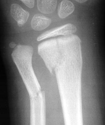
     This radiograph shows a fracture of the distal ulna
and radius.  The radius fracture extends from the
metaphysis into the physis.  The physis appears to
be slightly widened consistent with SH-II.

     An SH-III fracture occurs through the physis and
epiphysis (E).  Since this fracture often involves the
articular surface, this injury is more prone to chronic
disability if anatomic realignment is not achieved.

See SH-III example.

     This radiograph shows a fracture of the distal tibia
over the articular surface into the epiphysis and physis.

     An SH-IV fracture is a contiguous fracture through
the metaphysis, physis, and epiphysis.  This fracture
often involves the articular surface, making this a
high-risk injury for chronic disability as in SH-III
injuries.

See SH-IV  example.

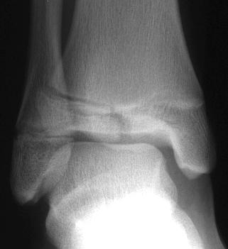
     This radiograph shows a fracture of the medial
malleolus extending from the inferior articular surface of
the tibial epiphysis through the physis and extending
through the metaphysis.

     An SH-V fracture is a crush injury of the physis.
This may be radiographically visible as a narrowing of
the growth plate lucency; however, it is most often not
radiographically visible.

See SH-V example.

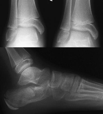
     This patient fell off a second story balcony onto
her feet.  The radiographs show several fractures within
the body of the calcaneus.  A Salter-Harris type V injury
of the distal tibia  was suspected because of the
mechanism of injury.  However, this type of injury is
rarely visible on initial radiographs.  The injury must be
suspected clinically.  Subsequent growth arrest of this
area confirms the presence of the Salter-Harris type V
injury.
     Comparison views of the non-affected extremity may
assist in radiographically diagnosing a SH-V type injury
at initial presentation.  Based on this comparison view,
differences in the width of the growth plates may be
evident.  A complete obliteration or diminished physeal
distance of the affected extremity confirms the
diagnosis of a SH-V injury.   However, even if there are
no obvious differences on the comparison view, or if a
comparison view is not obtained, or if both extremities
are injured, the patient should be treated as a possible
SH-V injury if the mechanism of injury suggests an axial
compression along the long axis of the bone, and the
patient exhibits tenderness along the physeal region.

References
     1.  Bachman D, Santora S.  Orthopedic Trauma.
In:  Fleisher GR, Ludwid S (eds).  Textbook of
Pediatric Emergency Medicine, third edition.
Baltimore, MD, Williams and Wilkins, 1993,
pp. 1237-1238.
Return to Radiology Cases In Ped Emerg Med Case Selection Page
Return to Univ. Hawaii Dept. Pediatrics Home Page
 An SH-I fracture may be suspected on clinical
grounds alone. The fracture line may not be
radiographically evident if the epiphysis is not displaced.
Tenderness over the physis should lead you to suspect
an occult SH-I fracture in the region of the tenderness
even if the radiographs are normal. This commonly
occurs in a wrist injury where normal wrist radiographs
may lead one to the pitfall of diagnosing a wrist sprain.
If there is tenderness over the physis of the distal radius
or ulna, a clinical diagnosis of an SH-I fracture of this
area should be made. An SH-I fracture is only visible
radiographically if the physis is widened, distorted, or
the epiphysis is displaced.
Radiographic confirmation of clinical SH-I fractures
may be made later during orthopedic follow-up by
stress views or the presence of new bone formation
along the physis approximately 7-10 days post-injury.
See SH-I example
 This radiograph shows a tiny fracture of the ulnar
styloid. The AP view is otherwise unremarkable. The
patient had point tenderness over the dorsum of the
radial physis. The lateral view shows a displaced radial
epiphysis. On careful inspection, you can see that the
radial epiphysis is not centered over the metaphysis.
The radial epiphysis is slightly displaced dorsally with
respect to the metaphysis. No fractures of the
epiphysis or the metaphysis are visible. Since the
fracture is strictly through the physis, this is a
Salter-Harris type I fracture.
An SH-II fracture occurs through the physis and
metaphysis (M).
See SH-II example
 This radiograph shows a fracture of the distal ulna
and radius. The radius fracture extends from the
metaphysis into the physis. The physis appears to
be slightly widened consistent with SH-II.
An SH-III fracture occurs through the physis and
epiphysis (E). Since this fracture often involves the
articular surface, this injury is more prone to chronic
disability if anatomic realignment is not achieved.
See SH-III example.
 This radiograph shows a fracture of the distal tibia
over the articular surface into the epiphysis and physis.
An SH-IV fracture is a contiguous fracture through
the metaphysis, physis, and epiphysis. This fracture
often involves the articular surface, making this a
high-risk injury for chronic disability as in SH-III
injuries.
See SH-IV example.
 This radiograph shows a fracture of the medial
malleolus extending from the inferior articular surface of
the tibial epiphysis through the physis and extending
through the metaphysis.
An SH-V fracture is a crush injury of the physis.
This may be radiographically visible as a narrowing of
the growth plate lucency; however, it is most often not
radiographically visible.
See SH-V example.
 This patient fell off a second story balcony onto
her feet. The radiographs show several fractures within
the body of the calcaneus. A Salter-Harris type V injury
of the distal tibia was suspected because of the
mechanism of injury. However, this type of injury is
rarely visible on initial radiographs. The injury must be
suspected clinically. Subsequent growth arrest of this
area confirms the presence of the Salter-Harris type V
injury.
Comparison views of the non-affected extremity may
assist in radiographically diagnosing a SH-V type injury
at initial presentation. Based on this comparison view,
differences in the width of the growth plates may be
evident. A complete obliteration or diminished physeal
distance of the affected extremity confirms the
diagnosis of a SH-V injury. However, even if there are
no obvious differences on the comparison view, or if a
comparison view is not obtained, or if both extremities
are injured, the patient should be treated as a possible
SH-V injury if the mechanism of injury suggests an axial
compression along the long axis of the bone, and the
patient exhibits tenderness along the physeal region.
References
1. Bachman D, Santora S. Orthopedic Trauma.
In: Fleisher GR, Ludwid S (eds). Textbook of
Pediatric Emergency Medicine, third edition.
Baltimore, MD, Williams and Wilkins, 1993,
pp. 1237-1238.