Drooling, Stridor, and a Barking Cough: Croup??
Radiology Cases in Pediatric Emergency Medicine
Volume 1, Case 10
Rodney B. Boychuk, M.D.
Kapiolani Medical Center For Women And Children
University of Hawaii John A. Burns School of Medicine
An 18 month old female presented to the Emergency
Department with a history of fever, noisy breathing, a
harsh cough, and drooling. The fever and coughing
began yesterday, but tonight the fever is higher and the
cough sounds very harsh. The sound of this cough was
alarming to the parents. The highest temperature
measured was 39.5 degrees rectally. She was noted to
be drooling more than usual, but this was attributed to
teething. Her cry was more raspy than her normal cry.
She was not taking in solids well, but she was taking
liquids well.
Exam: VS T39.1 degrees rectally, P170, R28, BP
100/66. She appeared alert, awake, not toxic, in no
acute distress. She did not appear to prefer an upright
or a forward leaning position. Skin was warm & moist,
without rash. No head or sinus tenderness were noted.
Tympanic membranes were normal. The oral pharynx
was clear and the mucosa was moist. Excessive
drooling was not noticed by the examiner. The neck
was supple with small lymph nodes bilaterally. Heart
regular without murmurs. Lungs clear when resting.
However, when she was crying, mild inspiratory stridor
was noted. An occasional croupy cough was noted.
The abdominal exam was unremarkable. Color and
perfusion were good. A soft tissue lateral neck
radiograph was ordered.
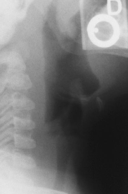
View lateral neck radiograph.
 Is this radiograph consistent with croup?
The epiglottis is normal in shape. The pre-epiglottic
(vallecular) space is preserved. The airway is patent.
There is pre-vertebral soft tissue swelling noted. This
radiograph is consistent with a retropharygeal abscess,
not croup.
Discussion and teaching points:
The retropharyngeal space is a pocket of connective
tissue that extends from the base of the skull
approximately to the tracheal carina. It harbors two
chains of lymphoid tissue that drain the nasopharynx,
adenoids, and posterior paranasal sinuses. Bacterial
infections of the areas drained may result in
suppuration of the nodes and abscess formation.
These lymphatic chains begin to atrophy about the third
or fourth year of life. Thus, 50% of the cases of
retropharyngeal abscess occur between 6 and 12
months of age, and 96% of cases occur in children
under 6 years of age (prior to lymphatic atrophy).
Staph aureus and group A beta-hemolytic streptococci
are the most common pathogens; however,
Hemophilus influenza and anaerobes have also
been recovered.
There is usually a prodromal nasopharyngitis or
pharyngitis with dysphagia, refusal of feeding, severe
throat pain, hyperextension of the head, and noisy
respirations. Previous trauma or evidence of
associated infectious conditions should be sought.
Respirations may be labored. There may be drooling,
stridor, a raspy voice (cry), and a croupy cough. A
bulge in the retropharynx may be visible. Meningismus
may result from irritation of the paravertebral ligaments.
Pain in the back of the neck or shoulder may be
precipitated by swallowing. However, in many cases, a
retropharyngeal abscess may be difficult to clinicially
distinguish from croup.
A lateral view of the soft tissues of the neck is
frequently helpful in making the diagnosis,
demonstrating the retropharyngeal mass in the stable
patient. Normal prevertebral spaces are as follows:
Anterior to C2: Less than or equal to 7mm in
children and adults.
Anterior to C3 and C4: less than 5mm in children or
adults or less than 40% of the AP diameter of the C3
and C4 vertebral bodies.
To simplify things, others suggest that the upper
pre-vertebral soft tissue should be no wider than one
vertebral body width.
Adequate hyperextension of the head and neck is
necessary in order to properly interpret the film if there
is no history of trauma. If the head and neck are not
properly positioned, the pre-vertebral space will appear
to be widened because the neck is not extended
enough. Repeating the radiograph with proper
positioning may resolve this problem. If proper
positioning is not possible or if the clinician is unsure if
plain films are definitive, CT of this area can more
accurately define any abnormalities of this region.
Most patients presenting with symptoms of croup
have viral croup. While epiglottitis is usually not
difficult to distinguish clinically from croup, an early
retropharyngeal abscess may be difficult to distinguish
from croup. A lateral neck radiograph may reveal this
occult diagnosis in selected cases, such as those with
high fever, unexpected lymphadenopathy, or those wit
h a suspicious bulge in the pharynx.
Other causes of partial upper airway obstruction
include epiglottitis, croup, peritonsillar abscess, severe
tonsillitis, infectious mononucleosis, cystic hygroma,
hemangioma, or neoplasms. Retained upper
esophageal foreign bodies, trauma to the retropharynx
from foreign body ingestion, instrumentation, and
C-spine injury can also cause localized swelling or
obstruction.
View another cause of stridor.
Is this radiograph consistent with croup?
The epiglottis is normal in shape. The pre-epiglottic
(vallecular) space is preserved. The airway is patent.
There is pre-vertebral soft tissue swelling noted. This
radiograph is consistent with a retropharygeal abscess,
not croup.
Discussion and teaching points:
The retropharyngeal space is a pocket of connective
tissue that extends from the base of the skull
approximately to the tracheal carina. It harbors two
chains of lymphoid tissue that drain the nasopharynx,
adenoids, and posterior paranasal sinuses. Bacterial
infections of the areas drained may result in
suppuration of the nodes and abscess formation.
These lymphatic chains begin to atrophy about the third
or fourth year of life. Thus, 50% of the cases of
retropharyngeal abscess occur between 6 and 12
months of age, and 96% of cases occur in children
under 6 years of age (prior to lymphatic atrophy).
Staph aureus and group A beta-hemolytic streptococci
are the most common pathogens; however,
Hemophilus influenza and anaerobes have also
been recovered.
There is usually a prodromal nasopharyngitis or
pharyngitis with dysphagia, refusal of feeding, severe
throat pain, hyperextension of the head, and noisy
respirations. Previous trauma or evidence of
associated infectious conditions should be sought.
Respirations may be labored. There may be drooling,
stridor, a raspy voice (cry), and a croupy cough. A
bulge in the retropharynx may be visible. Meningismus
may result from irritation of the paravertebral ligaments.
Pain in the back of the neck or shoulder may be
precipitated by swallowing. However, in many cases, a
retropharyngeal abscess may be difficult to clinicially
distinguish from croup.
A lateral view of the soft tissues of the neck is
frequently helpful in making the diagnosis,
demonstrating the retropharyngeal mass in the stable
patient. Normal prevertebral spaces are as follows:
Anterior to C2: Less than or equal to 7mm in
children and adults.
Anterior to C3 and C4: less than 5mm in children or
adults or less than 40% of the AP diameter of the C3
and C4 vertebral bodies.
To simplify things, others suggest that the upper
pre-vertebral soft tissue should be no wider than one
vertebral body width.
Adequate hyperextension of the head and neck is
necessary in order to properly interpret the film if there
is no history of trauma. If the head and neck are not
properly positioned, the pre-vertebral space will appear
to be widened because the neck is not extended
enough. Repeating the radiograph with proper
positioning may resolve this problem. If proper
positioning is not possible or if the clinician is unsure if
plain films are definitive, CT of this area can more
accurately define any abnormalities of this region.
Most patients presenting with symptoms of croup
have viral croup. While epiglottitis is usually not
difficult to distinguish clinically from croup, an early
retropharyngeal abscess may be difficult to distinguish
from croup. A lateral neck radiograph may reveal this
occult diagnosis in selected cases, such as those with
high fever, unexpected lymphadenopathy, or those wit
h a suspicious bulge in the pharynx.
Other causes of partial upper airway obstruction
include epiglottitis, croup, peritonsillar abscess, severe
tonsillitis, infectious mononucleosis, cystic hygroma,
hemangioma, or neoplasms. Retained upper
esophageal foreign bodies, trauma to the retropharynx
from foreign body ingestion, instrumentation, and
C-spine injury can also cause localized swelling or
obstruction.
View another cause of stridor.
 This radiograph shows evidence of epiglottitis (also
called supraglottitis). The epiglottis is thumb-like in
appearance (instead of triangular or flat in shape) and
the aryepiglottic folds are thickened. The pre-epiglottic
space is preserved to some degree, but it is not as
large as it should be. In many cases of epiglottitis, the
pre-epiglottic space is obliterated (replaced by
edematous tissue). The retropharyngeal space
(pre-vertebral tissue) is not widened.
View another cause of stridor.
This radiograph shows evidence of epiglottitis (also
called supraglottitis). The epiglottis is thumb-like in
appearance (instead of triangular or flat in shape) and
the aryepiglottic folds are thickened. The pre-epiglottic
space is preserved to some degree, but it is not as
large as it should be. In many cases of epiglottitis, the
pre-epiglottic space is obliterated (replaced by
edematous tissue). The retropharyngeal space
(pre-vertebral tissue) is not widened.
View another cause of stridor.
 This radiograph looks normal except for a mild
degree of subglottic airway narrowing. This type of
pattern correlates best with patients presenting with
viral croup.
References
Fleisher GR. Infectious Disease Emergencies. In:
Fleisher GR, Ludwig S (eds). Textbook of Pediatric
Emergency Medicine, third edition. Baltimore, Williams
& Wilkins, 1993, pp. 613-621.
Santamaria J, Abrunzo TJ. Ear, Nose, and Throat.
In: Barkin R (ed). Pediatric Emergency Medicine
Concepts and Clinical Practice. Chicago, Mosby Year
Book, 1992, pp. 680-682.
This radiograph looks normal except for a mild
degree of subglottic airway narrowing. This type of
pattern correlates best with patients presenting with
viral croup.
References
Fleisher GR. Infectious Disease Emergencies. In:
Fleisher GR, Ludwig S (eds). Textbook of Pediatric
Emergency Medicine, third edition. Baltimore, Williams
& Wilkins, 1993, pp. 613-621.
Santamaria J, Abrunzo TJ. Ear, Nose, and Throat.
In: Barkin R (ed). Pediatric Emergency Medicine
Concepts and Clinical Practice. Chicago, Mosby Year
Book, 1992, pp. 680-682.
Return to Radiology Cases In Ped Emerg Med Case Selection Page
Return to Univ. Hawaii Dept. Pediatrics Home Page