Succineid Morphology |
||||
|
We have undertaken anatomical analysis of 19 Hawaiian species of succineids. Data collection and analysis of conchological characters has been carried out for over 200 specimens representing a variety of Hawaiian taxa, including type specimens from the collections at the Bishop Museum in Honolulu. Soft
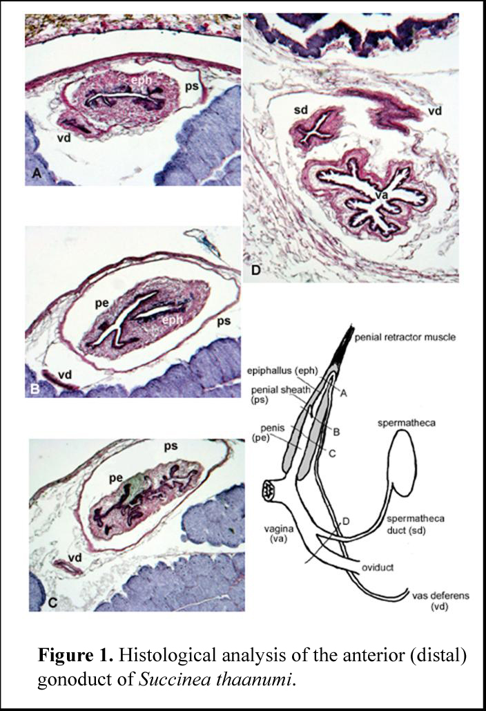
Anatomy Figure 1
illustrates one of the primary character sets, the distal reproductive
system, in this case of Succinea
thaanumi from the
Figure 1B
shows a cross-section through the posterior region of the penial sheath,
where the epiphallus and penis are separated. The penis and epiphallus are
both muscular. The penis has a Y-shaped lumen, and the epiphallus at this
point has a simple lumen with some mucoidal secretory cells. The vas
deferens is a thin-walled duct to the lower left. An interesting
characteristic (perhaps species-specific) is the extension of the penial
lumen past its junction with the epiphallus. Figure 1D
is more ventral in the body, and is a section through the vagina, or
anterior oviduct. The oviduct itself is deeply folded without a thick
layer of muscle, and the lumen is nonglandular. The spermathecal duct,
x-shaped in cross-section, is above left, and the vas deferens, cut on an
angle, is above right. |
||||
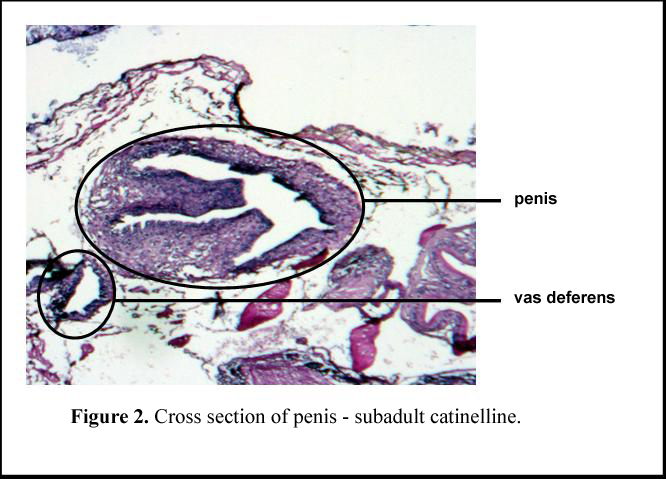
| Analysis of the anatomical data indicate that they are consistent with molecular data in recognizing two major clades, corresponding to the two traditional subfamilies Catinellinae and Succineinae. These are differentiated on the basis of presence or absence of a penial sheath. Catinellines lack the penial sheath (Figure 2), whereas succineines have an external sheath around the penis, creating a tubular introvert within a sac (Figure 3). | ||||
 |
 |
|||
|
Organogenetic patterns may provide a clue as to how these two morphologies diverged, by illustrating developmental similarities between them. Results show that the succineid gonoduct, like those of other pulmonates, develops as a singular structure that originates with the anterior common genital opening, and extends posteriorly as it grows and develops. This is unlike the reproductive systems of more primitive marine gastropods, which develop from two or more primordia and essentially ‘meet in the middle’. The succineid species we have investigated mature relatively late in growth, and the gonoduct is typically not developed completely until the individual is almost full grown. The difference between the two subfamilies is present relatively early in development of the structure. Half-grown individuals representing both taxa have their subfamilial characteristics already present, although the structures are not fully developed.In Figure 2 (subadult catinelline) the penis and vas deferens are present and, though not developed, are anatomically recognizable as being of the catinelline clade type. The penis lacks a sheath and the internal penial duct is even at this stage fairly complex. In the subadult succineine (Figure 3), the thin walled sheath of the penis is present around the outside of the structure, and the lumen of the penis itself is relatively simple. There are two possible interpretations of these results. The catinelline penis may be homologous to the succineine penis without the sheath, such that the succineine penial sheath is an autapomorphy. Alternatively, the succineine penis sheath may be homologous to the catinelline penis, in which case the internal folds of the catinelline structure (e.g., the two large folds that run the length of the penis on the left side in Figure 2) may have disconnected to form a separate tubular structure. One
species in the Hawaiian catinelline clade has anatomical characteristics
suggesting placement in the catinelline genus Quickia (all the rest investigated in that clade have anatomical
character states consistent with placement in Catinella); further analysis of additional species will be needed to
determine whether Quickia is in
fact present in Hawaii, or whether the character states that suggest that
placement are homoplastic. |
||||
|
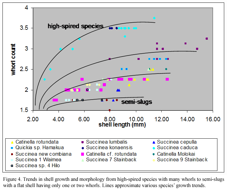
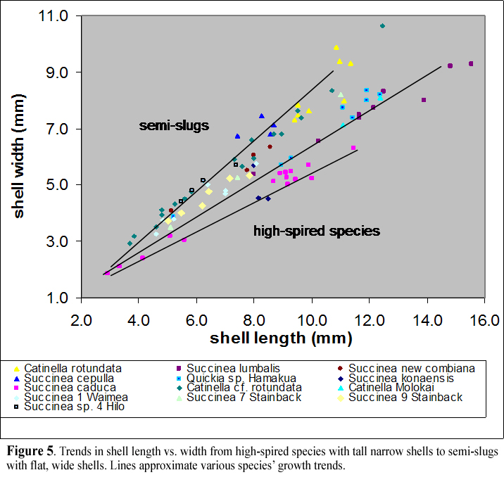
Shell Morphology Many succineid shells are highly reduced, and they vary relatively little in many respects among species. However, shell shape is one of the primary morphological characters we are interested in with this study, because it does vary in succineids between low-spired, very flat wetland species and higher spired, more xeric species. Shell length, aperture length, shell width and whorl numbers have been measured to tease out these aspects. Within the two major groups, Succineinae and Catinellinae, species have evolved convergently along a range of shell morphologies (Figure 4, Figure 5), such that both groups include very flat-shelled slug-like species (e.g. Succinea newcombiana), as well as typical intermediate forms. The succineine clade also includes at least two high-spired species found in relatively arid habitats (Succinea caduca and Succinea konaensis). |
||||
 |
 |
|||
|
Radula Morphology Other hard parts of mollusks also provide useful characteristics; in succineids, the jaw and the radula or teeth have been reported for most species and contain some useful information. The jaw is similar morphologically across all species noted. They vary somewhat in the strength of development of the center point of the jaw, which in some Succinea species from the island of Hawaii is essentially absent. The radular teeth are very similar among species; the radula has a symmetrical rachidian tooth, and mitten-shaped lateral teeth with single large cusps and flanking smaller cusps. Mollusca n radular tooth fields are typically divided into a center rachidian tooth, lateral teeth flanking the rachidian tooth, and marginal teeth toward the outside. In the species investigated, there is very little variation in tooth shape; the boundary between lateral and marginal teeth is placed at the point where the single large cusp evident in the rachidian and inner teeth divides into two or more smaller cusps. The location of the boundary between tooth fields is thus somewhat arbitrary, and it varies somewhat within species. Interestingly, in the species investigated, the number of lateral teeth appears to increase more through ontogeny than the number of marginals. Succineid species tend to vary in relative numbers of teeth in each field; catinellines and some succineines have about four times as many marginal as lateral teeth, whereas other succineines have about equal numbers of marginal and lateral teeth. Individuals as adults typically have about 70 to 130 rows of teeth. |
||||